What Does a Newborn Baby's Chest Look Like
Abstract
The highly compliant nature of the neonatal chest wall is known to clinicians. All the same, its morphological changes have never been characterized and are especially important for a customised monitoring of respiratory diseases. Hither, nosotros show that a device applied on newborns tin trace their breast boundary without the utilize of radiation. Such engineering science, which is easy to sanitise betwixt patients, works like a smart measurement tape drawing also a digital cantankerous department of the chest. We also prove that in neonates the supine position generates a significantly dissimilar cross section compared to the lateral ones. Lastly, an unprecedented comparison between a premature neonate and a kid is reported.
Introduction
Newborns featuring lung immaturity require continuous monitoring and treatment in Neonatal Intensive Care Units (NICU); an intervention that is critical for the survival of preterm babies. An estimated fifteen 1000000 premature neonates are born each year1. Recently, the CRADL project has introduced the continuous assessment of regional lung function using Electrical Impedance Tomography (EIT) technology as supportive care for the most common causes of paediatric respiratory failure (http://cradlproject.org/). As part of the projection, a textile electrode patient interface for the neonatal EIT measurement (SenTec AG, CH) has been adult and tested to validate its clinical operation in a multicentre clinical written report2. Results showed the absence of any discomfort for the patients whilst providing a low contact impedance, which ensures that good quality measurements can be taken. Hence, such technology gives clinicians a novel insight about the upshot of the prescribed therapies on newborns' respiration3,iv. However, it has been noticed that in some patients the fitting of the device, designed as a chugalug for the thorax, was non optimal in all lying positions of the babies. The highly compliant chest wall of neonates increases the tendency to chest wall recession specially for premature babies5. Thus, respiratory diseases leading to poor lung compliance makes neonates prone to respiratory failure and the need for mechanical ventilation. Therefore, an investigation almost the geometric alter of the chest wall was launched every bit role of the CRADL project, aiming to amend the performance of the device.
Measurements of the newborn chest are not office of the consolidated clinical routine. At birth, the height, the weight and the head circumference are noted by routine. Therefore, to the best of the authors' knowledge, no study has collected data about the chest perimeter of newborns and its physiological biomechanical behaviour.
Such data is critically important for predicting the plumbing fixtures of the neonatal belt in patients of different weight and gestational ages. Almost a century agone, Schultz6 pointed out that the literature on homo foetal growth was not very extensive. Present, whilst it is well known by clinicians that the morphology of the human chest changes significantly from the neonatal to the adult stages, no extant database quantifies this evolution. In the 80s, a rare chest morphology written report compared the influence of age and sex on loftier altitude populations past conveying out anthropometric measurements at the end of a normal expirations in the age range v–24 years old7. Recently, Chang et al.8 accept presented a non-invasive method to record the shape of the anterior chest wall to care for infants affected by pectus excavatum. Although nowadays chest CT and radiographs provide the required details, children are much more radio-sensitive than adults. Hence, they decided to attach a strip of thermal plastic to obtain a permanent profile of the inductive chest wall8. The strip solidified after 10 providing a cross-exclusive view of the thorax, which was then scanned for subsequent data analysis.
Therefore, there is a clinical need to acquire, without the use of radiation, the torso shape for treating a diverseness of respiratory diseases, which are particularly critical for infants. The authors have recently presented a vesture device featuring accelerometers that could successfully carry out the acquisition of unlike shape boundaries in vitroix.
The nowadays study aims at applying the developed technology in vivo for an unprecedented collection of neonatal torso shape related measurements performed in real time past means of an electronic measuring belt. Hence, the goal is to quantify, by means of upward-to-date technology, the biomechanical changes of the neonatal chest in different lying positions. As a consequence, the morphological differences observed in a small population of newborns volition be available to neonatologists. Such insight will also give a better understanding most the different configurations that any EIT device needs to adapt to. Therefore, the overall objective is to empathize how the mechanical aspect of EIT monitoring could be improved for an increased quality of data supplied to clinicians.
Results
The novel electronic measuring belt was used on 1 child and 31 newborns, 6 of which were in the NICU. Information technology should be noted that patient no. 20 could not be placed in the prone configuration because of a tracheotomy. Similarly, patient no. 31 could non be measured in the prone position as she was intubated at the time of the study, hence information technology has not been possible to acquit out the complete protocol.
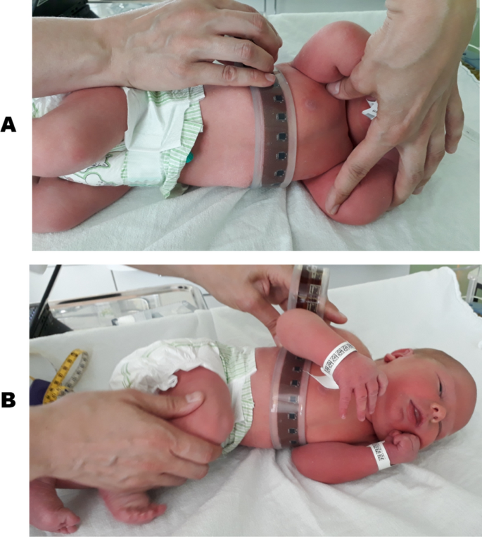
The electronic measuring belt practical on one of the recruited subjects in the motherhood ward. According to the protocol, in this shot the clinician was recording the body shape of the babe lying on his left side with his artillery up (A) and downwards (B). The photographs are published with permission of both of the infant's parents.
None of the measurements were interrupted because of the device or because the parents attending the report lacked conviction in the process carried out on their babies. No restriction of breathing or skin irritation was observed. The belt was laid on the mattress while each baby was placed on top of information technology perpendicular to the chugalug. The belt was wrapped effectually the breast and held in place past the clinician, as shown in Fig. 1, for less than 5 due south whilst the measurement was captured. In society to modify the position, the belt was unwrapped, the baby rotated to the next required position and the chugalug wrapped into position in one case once again for the next measurement to be captured. The study was conducted at a baby-friendly pace, meaning that sleepy participants were faster to move around whilst the more agile ones were offered more fourth dimension to settle in the position. Overall, the written report took no more than 5 min to be completed on each bailiwick. The data of the recruited subjects are listed in Table one. Over 80% of the newborns were born at term, existence defined subsequently 37 weeks of Gestational Historic period. 16 of the newborn participants were male and fifteen were female.
The performance of the belt has been analysed by computing its accuracy and repeatability on the recruited newborns. Since the tape measurement was performed on all subjects in the supine position, this was selected every bit the golden standard. Hence, the accuracy was calculated as the departure betwixt the mean of the two measurements carried out in supine position and the chest perimeter reported in Table one. The maximum offset of i.7 cm was reported for subject number ix, whilst the minimum of 0.5 mm was estimated for patient no. ten. Overall the average accuracy was 5.7 mm. The repeatability was determined as the difference in modulus between the two measurements in the supine position for the same patient recorded while conveying out the protocol. As a result, the range is between 0.2 mm for patient no. 20 and 1.5 cm for patient no. 7, whilst the average repeatability is six.vi mm.
The perimeter, the area and the size part of the curve describing the torso cross-section accept been calculated in every recorded position for each patient. Table 2 reports the boilerplate of these values in the supine position, which was recorded twice during the protocol. In a preliminary analysis the effect of the gestational historic period (hence the weight) was neglected and the lying position in which the minimum and maximum for each of the above three features were recorded for each subject was determined. As a result, the minimum circumference is estimated when the babe was lying on the right side for the majority of the subjects (32%), whilst the maximum was recorded for the babe lying supine (35%). A similar trend is captured for the area, beingness minimum mostly on the right side (29%), whilst the maximum is expected when the infant is 45 degrees to the right (26%) followed by the supine position (23%). An coordinating outcome is obtained for the size part, which is minimum for the same number of patients lying 45 or ninety degrees to the right (29%) and maximum in the supine position (77%).
In society to identify meaning thoracic changes, statistical analyses have been carried out on the entire population and on a selection of four different gestational ages: 30 weeks, 33 weeks, 39 weeks and 41 weeks. The first ii characterise the biomechanics of early on pre-term babies whilst the latter two define the term subjects.
The Anderson–Darling test established that the breast circumference and area values practise not come up from a normal distribution. Hence, the Kruskal–Wallis exam has been adopted to determine if the samples come from the same population. Firstly, dividing the population into just two groups, preterm (less than 37 weeks of gestation) and term, highlights a considerable discrepancy in both the circumference and surface area. The choice based on the weeks of gestation is justified as the early on pre-term subjects (30 weeks, 33 weeks) show anatomical dimensions significantly different from subjects built-in after 39 weeks of gestation (p values beingness 2.69e−06 for perimeter and 3.3e−06 for expanse). As the main interest of this study consists in identifying peculiar changes in the newborns' chest acquired by their lying position, four accept been selected from the protocol: prone, 90 degrees to right, 90 degrees to left and supine. This selection is a event of the trends shown previously among all included subjects and aims to identify the main biomechanical changes. As an case the chest boundaries computed for patient no. 30 in different positions are shown in Fig. two. Considering the position every bit unique grouping variable fails to notice whatever significant change in the circumference and area.
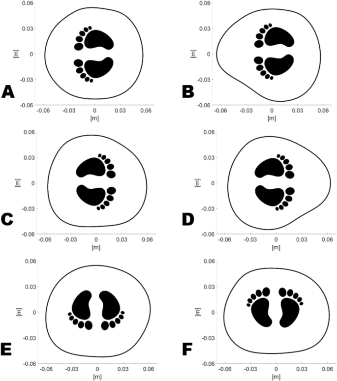
Comparison of the computed breast boundaries for subject area no. 30. The babe was lying on her right side with her arms upwardly (A) and down (B), on her left with her arms up (C) and downwardly (D), prone (E) and supine (F).
On the other hand, the computed size function values are normally distributed. Thus, the ANOVA examination were performed. In this case the event of the supine position alone generates shapes significantly different from lying both on the correct and on the left side (p is seven.6e−05). Similarly, the prone position leads to statistically distinctive shapes compared to the lateral ones (p is 1.7e−04). Comparing only the prone to the supine highlights another substantial difference (p is 0.017), which is not evident in one case the iv configurations are confronted.
A further multiway ANOVA investigated in detail the issue of the position and the week of gestation on the size function. As a result, for each of the selected week of gestation the values on the right are never statistically different from the ones on the left. Lying on the right side though produces shapes significantly different than the supine position for the subjects built-in at 39 weeks of gestation.
In order to dominion out the effect of other factors on the observed change of chest shape, statistical analyses were carried out to test also the sex of the subjects as an contained variable. As a result, the sex is leading to no pregnant changes in terms of chest perimeter, area or size function even for the subjects built-in at 39 weeks of gestation. Lastly, testing for the issue of the ward where the subjects were cared for has not been thought sensible equally all pre-term babies and only i newborn born at 39 weeks were in the NICU. Hence, no statistical consideration would be significant.
Discussion
An unprecedented data drove about the purlieus changes in the neonatal torso due to the lying position has been presented in this report. Although it is known to neonatologists that the chest of newborns is very compliant, no characterization was available. This miracle is highly relevant to the awarding of a patient interface EIT belt to monitor the lungs' ventilation in newborns. The patient interface EIT belt in the CRADL project was suffering from loose plumbing equipment, with associated loss of electrode contact, in some positions for a number of subjects, hence the evolution of an electronic shape measuring belt for this investigation, which included 31 newborns and 1 kid.
The accurateness (mean v.7 mm) and repeatability (mean 6.6 mm) of the measuring belt take been assessed and judged satisfactory. It is worth noticing that the thickness of the encapsulation has been neglected. The thickness of the silicone rubber between the skin of the baby and the printed circuit board, where the accelerometers are placed, is ii mm, which is controlled past the mould depth. Platinum cure silicone was used, which does not exhibit cure shrinkage. Even so, given the bespoke paw-made process information technology is difficult to establish whatsoever tiny dimensional change in this layer and to estimate the actual boilerplate thickness alter. In add-on, no compression examination was carried out to quantify the deformation of such silicone encapsulation under the average weight of the subjects.
Figure two shows the breast boundaries, reconstructed in different positions, as an example of what happens in a typical term newborn. Firstly, the chest appears slightly compressible every bit the cross-exclusive area is maximum in the prone position, decreases of iv.5% in the supine position and of 7% when lying ninety degrees to each side for this bailiwick. Hence, it appears that the compression of the ribs, quite flexible at this age, plays a big role in the biomechanics of the torso. The distortion of the chest wall has been attributed to its increased compliance compared to the lung, to the incomplete ossification of the ribs and to the respiratory muscles unable to stabilize the chest wall10. Infants feature horizontally placed ribs compared to the downward-facing ones in adults, which let a significant increase in both the anteroposterior and lateral diameters of the thorax when the diaphragm descends5. Nigh ii years are needed for the infant rib-cage to mature into the adult configuration, stabilized by the external intercostal muscles, hence the animate patterns compensate for the anatomic immaturity5.
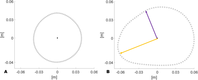
Comparison of the computed chest boundaries which feature the minimum (A) and the maximum (B) value of shape function. The purple arrow indicates the minimum altitude from the centroid, whilst the orange one is the maximum altitude.
A visible alter in the morphology of the chest cross section is presented, especially when the lower arm is compressing the chest (Fig. 2B,D) compared to the other positions. The size function has been introduced to quantify and differentiate the shapes recorded in a way that the higher the value, the more irregular is the shape. In other words, the lower its value the more circular the shape looks similar every bit shown in Fig. 3. The breast boundary of patient no. 19 (Fig. 3A) lying on his left side, being the lightest in weight amid the recruited patient, looks almost like a regular circle. Differently, the boundary of patient no. 7 (Fig. 3B) lying supine appears disproportionate every bit the altitude from the centroid ranges from 4.45 cm (purple arrow) to 6.66 cm (orangish arrow).
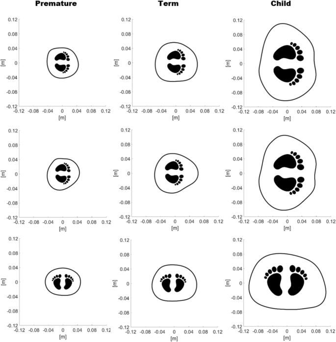
Development of the torso boundary computed for patient no. 31 (premature), patient no. 30 (term) and a child. On the elevation row the subjects are lying on their left side without compressing the torso with their arm. On the middle row their left arm lies between the mattress and the chest. On the lesser row the study subjects are lying supine.
One of the artillery normally compresses the body when lying on a side as newborns practise not heighten them. Hence the statistical analyses accept taken into account only this physiological configuration.
An overall trend has been observed as the majority, approximately 30% of the subjects, exhibited minimum values of chest perimeters and area when lying on the correct side. This issue could be explained by the fact that the highly compliant chest wall easily compresses more the right side, which unlike the left one does non feature the heart. Thus, the hypothesis emerges that the heart itself slightly constrains the deformation of the left side. The size function highlighted that a maximum value is observed in the supine position for 77% of the subjects, significant that a greater scatter between the anteroposterior and lateral diameters leads to a more oval cross department.
The Anova test confirmed that because only the event of the four main configurations (prone, 90 degrees to correct, ninety degrees to left and supine) the size functions computed are statistically distinctive comparing each of the lateral configurations to the prone or the supine ones. In addition, dividing the population into preterm and term and comparing the iv configurations, the supine position is significantly unlike from the lateral ones only in the term subjects. A further Anova enquired if such difference is significant also in each group of gestational age. Equally a result, a remarkable divergence betwixt the size functions obtained on the right side compared to the supine position is estimated for the subjects born at 39 weeks of gestation. Notwithstanding, such a event could be biased by the highest number of samples (8 subjects) for the babies born later on 39 weeks compared to the other subgroups. It is worth remembering that the nowadays preliminary investigation is not a clinical report, thus the limited sample size. Notwithstanding, it has been noticed that the trunk purlieus deforms diversely in newborns based on their gestational age, thus peradventure considering of the weight. The preterm subjects (30 and 33 weeks) exhibit a less pronounced deformation of the chest in one case placed on their side. Such an effect appears to be in contrast with the high compliance of the neonatal chest wall. However, these subjects feature an boilerplate weight of 2.042 kg confronting the mean of 3.524 kg at 39 weeks. Given the lower mass, a lower force is practical to the organs of the preterm babies, which module leads to express deformations.
This work was able to test the measuring belt also on a child of 20 kg aged 6 years. The boundary obtained is compared to a premature newborn and a term one in Fig. 4. This analogy is novel, to the best of the authors' knowledge, every bit it shows the maturation of the chest in young infants. On the tiptop row it is clear that the sternum and the spine are visible only in the kid. In the middle row, it is shown that the arm placed betwixt the mattress and the chest generates a more pronounced deformation of the cantankerous section in the term newborn. Such effect could exist explained by the different weight, being 1.795 kg for the premature and 4.355 kg for the term, if compared with the other neonatal breast and by the different ossification if compared to the child. Finally, on the bottom row, although the dimensions are diverse for each subject, the boundary shapes appear comparable as gravity leads to an increased lateral bore and to the disappearance of the concavity on the sternum fifty-fifty for the child.
This preliminary investigation intends to report an insight into the evolution of the chest in the infants and to demonstrate how the thorax changes in neonates. This information could be used by clinicians in medical do, by engineers and designers in order to take into account such changes to improve the EIT belt fitting and to differentiate the reconstruction algorithm, which at the moment, to the all-time of the authors' cognition, neglects any deformation of the breast or pinch of the lungs.
Methods
Study population and protocol
This study was carried out within the framework of the CRADL project, which has received funding from the European Spousal relationship's Horizon 2020 enquiry and innovation programme. Post-obit the approval of the Ethical Committee of the Northern Ostrobothnia Wellness Intendance District (ETTMK: 60/2018) 34 babies were asked to participate, three of whom the parents declined (1 in maternity ward, 2 in NICU). The study was performed in accordance with the relevant guidelines and regulations. Later obtaining written informed consent of both parents, 31 newborns and i child were included in total from the Oulu University Infirmary, Oulu, Finland. Informed consent from both parents was obtained for publication of the photographs in an online open-admission publication.
At the beginning of the protocol, neonates were positioned supine on the mattress of an open incubator, above which the radiant warmer was already switched on for their comfort. Parents were welcomed to observe the whole process, which was carried out by the paediatricians amongst the authors of this written report. Although statistically not relevant, the child was included in the report aiming to gain a preliminary insight about the thoracic change compared to the newborns and to assess the performance of the device on an older subject. The following details were acquired for each patient:
-
Gestational historic period in weeks and days.
-
Postnatal age in hours, weeks and days.
-
Sex.
-
Ward: maternity or NICU.
-
Acme at birth in cm.
-
Weight at nascence in cm.
-
Head circumference at birth in cm.
-
Chest circumference at the time of the study in cm.
The Breast circumference was evaluated below the nipple line by means of the standard measurement tape, similarly to what is done at birth for the Head. Successively, the electronic measuring belt was used to perform 7 measurements of the torso in 6 dissimilar positions as follows:
- one.
supine;
- 2.
45 degrees to right;
- 3.
90 degrees to correct;
- 4.
prone;
- v.
45 degrees to left;
- vi.
ninety degrees to left;
- 7.
supine.
The repeated supine measurement was meant to assess the reliability of the records in comparing with the tape measure and to evaluate the sensitivity of the device.
The electronic measuring belt
The device is designed to piece of work every bit a smart tape. It features 32 high resolution three-axis accelerometers ADXL313 (Analog Devices, Norwood, MA, USA) every bit spaced every 16 mm. The electronics is perfectly insulated past means of a custom encapsulation made of a bio-compatible, platinum cure, medical course silicone condom, which prevents any possible current transfer to the skin of the babe. The encapsulation is soft, not-adhesive and has comfy rounded edges. The device can be easily sanitized for each patient by wipes usually used in the maternity wards. A prior soak examination and a thorough caption of the structure of the device was judged satisfactory past the ethical commission that approved the belt without additional safety testing. The belt was not classified equally a medical device. The accelerometers in the measurement belt are powered (3.3 Five) only by a laptop, sensors are then addressed and the data stored by an Arduino micro-controller connected to a laptop. The 32 accelerometers were assigned one of 2 I2C addresses and individually addressed via a 16 channel multiplexer (SparkFunElectronics, Boulder, CO, United states). The Arduino and multiplexer were housed in a transparent polycarbonate box (Hammond Manufacturing Ltd, Ontario, Canada). A live conquering data link was established between the Arduino and MatLab via the laptop serial connection. Data were acquired by Arduino at 0.five Hz and transmitted at 2 Mbps to the laptop. A command and visualisation interface was produced in MatLab to trigger the data collection, visualise the results and feedback on its completion. The belt is gently positioned around the chest and held in place by the clinician exactly similar a standard measuring tape with no fastening adopted.
The algorithm
Once the belt is positioned around the breast of each discipline, the measuring procedure is activated from the custom fabricated script in MatLab (Mathworks, Nantick, MA, U.s.) on the laptop via a serial communication. A flowchart is reported in Fig. five to show how the algorithm and the protocol have been integrated.
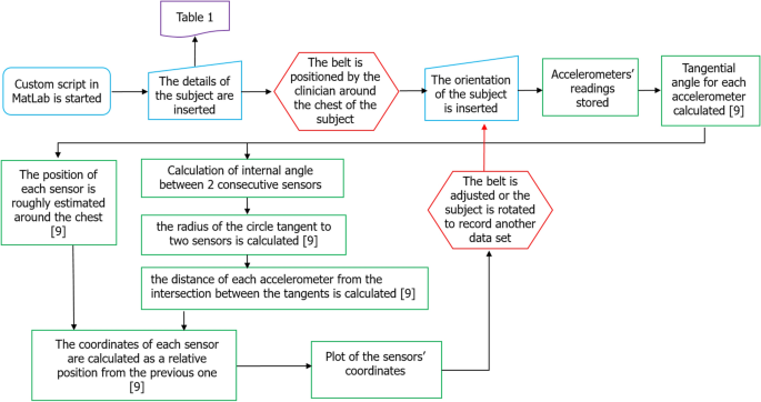
Algorithm steps (green) immune to double check the data acquisition during the protocol, which was carried out past a clinician (red) supported by an engineer (blue). A previous work9 explains the core of the algorithm.
The nuts of the algorithm needed to learn the torso shape via the accelerometers were reported in a previous worknine. A series of prompts were added to insert in real time the details listed in the above detailed protocol of each patient before the actual recordings. The tape of each position is instantaneously recorded, the cross-exclusive area plotted as feedback and the information saved on the laptop.
Every bit farther novelty to the published study9, a custom-made function was implemented to take into account that the belt was designed to exist long enough to accommodate the anatomical variability of all subjects. Therefore, the algorithm also automatically checks if there is a gap or overlap between sensor ane and sensor 32 by estimating the distance between them. The distance between all the other accelerometers is iteratively estimated and sensors overlapping for a distance below 15.ii mm were removed from the plot of the cantankerous-sectional breast area.
In case the infant suddenly moved or the chugalug was not positioned in a satisfactory configuration, further recordings were acquired. Information technology is important to note that the sensors on the belt were calibrated prior to the report by placing the chugalug on a rigid rig oriented in half dozen unlike configurations, accounting for the positive and negative components of the gravity vector recorded by the accelerometers forth the axes X, Y and Z.
Information assay
Once the relative location of the accelerometers effectively delineating the torso purlieus has been estimated, 100 equally spaced points in arc length were added past means of the interparc function in MatLab to obtain a improve fitting. The arc length interpolation was successively carried out past the interpclosed function, which returns the perimeter, the area and the centroid of the curve describing the torso cantankerous-section. Hence the coefficients of the piecewise polynomial interpolation are calculated and the area is estimated as an integral.
In order to quantify the modify in shape of the reconstructed cantankerous-sectional expanse, a size role has been introduced. The minimum and maximum distance from the centroid has been calculated for every recorded position. The size role is, therefore, defined as the difference between the maximum and the minimum of such distances (Fig. three).
The statistical analyses were carried out firstly by checking the assumption that all sample populations are normally distributed. Such an assumption has been checked by the Anderson–Darling test, implemented in MatLab equally adtest, which returns a test decision for the goose egg hypothesis. In instance the Anderson-Darling test fails to reject the zippo hypothesis at the default 5% significance level, the one-way Analysis of Variance (ANOVA), anova1, enables to find out whether different groups of an independent variable have different effects on the response variable. In the present report, the independent variable is, equally an example, the lying position of the infant, while the perimeter and expanse are the response variables. In order to test the event of multiple factors, such as the position and the gestational age, on the breast perimeter, area or size function a multiway ANOVA, anovan, has been employed. This test has been selected because of the unbalanced pattern, every bit the recruited subjects are not every bit distributed amidst the different gestational ages. In case the Anderson–Darling examination rejects the null hypothesis, the Kruskal–Wallis test, implemented in MatLab equally kruskalwallis, compares the medians of the groups of data to decide if the samples come from the aforementioned population.
References
-
WHO. Preterm Nascency (WHO, Geneva, 2018).
-
Sophocleous, Fifty. et al. Clinical performance of a novel fabric interface for neonatal chest electrical impedance tomography. Physiol. Meas. https://doi.org/10.1088/1361-6579/aab513 (2018).
-
Rahtu, M. et al. Early recognition of pneumothorax in neonatal respiratory distress syndrome with electrical impedance tomography. Am. J. Respir. Crit. Intendance Med. 200, 1060–1061. https://doi.org/ten.1164/rccm.201810-1999IM (2019).
-
Kallio, Grand. et al. Electrical impedance tomography reveals pathophysiology of neonatal pneumothorax during NAVA. Clin. Instance Rep. 8, 1574–1578. https://doi.org/10.1002/ccr3.2944 (2020).
-
Stocks, J. & Hislop, A. Structure and function of the respiratory organization: Developmental aspects and their relevance to aerosol therapy. In Drug Delivery to the Lung (eds Bisgaard, H. et al.) (Taylor & Francis Group, New York, 2001).
-
Schultz, A. H. Fetal growth of man and other primates. Q. Rev. Biol. 1, 465–521. https://doi.org/ten.1086/394257 (1926).
-
Beall, C. A comparison of chest morphology in high altitude Asian and Andean populations. Hum. Biol. 54, 145–163 (1982).
-
Chang, P. Y. et al. A method for the non-invasive assessment of chest wall growth in pectus excavatum patients. Eur. J. Pediatr. Surg. xx, 82–84. https://doi.org/10.1055/s-0029-1241819 (2010).
-
De Gelidi, S. et al. Trunk shape detection to meliorate lung monitoring. Physiol. Meas. https://doi.org/10.1088/1361-6579/aacc1c (2018).
-
Davis, Thousand. M., Coates, A. 50., Papageorgiou, A. & Bureau, M. A. Direct measurement of static chest wall compliance in animal and human being neonates. J. Appl. Physiol. 65, 1093–1098. https://doi.org/10.1152/jappl.1988.65.three.1093 (1988).
Acknowledgements
This work was supported in part by the CRADL project, funded by the European Spousal relationship's Horizon 2020 Enquiry and Innovation Programme 2014–2018 under Grant Agreement No. 668259, and the PNEUMACRIT project, and in part by the Engineering and Concrete Sciences Inquiry Council nether Grant No. EP/T001259/one. M.R. and M.G. are sponsored past the Finnish Foundation for Pediatric Research, Grant No. 190139. M.R. received also a personal research grant from The Alma and KA Snellman Foundation, Oulu, Finland. The authors are also grateful to the interns in RedLoop who helped design the custom mould needed for the encapsulation of the belt and the rig needed for its calibration. The icon adopted in the Figures is footprints by Kangrif from the Noun Project.
Author information
Affiliations
Contributions
A.B. and M.Chiliad. conceived the study, A.B. designed the belt, S.D. wrote the algorithm, Y.W. and A.D. designed the circuits of the belt, N.S. brash and encouraged to investigate the lateral positions, A.B., Due south.D., Chiliad.R. and M.K. conducted the acquisition, Southward.D. analysed the results and wrote the manuscript, R.B. supervised the projection. All authors reviewed the manuscript.
Corresponding author
Ethics declarations
Competing interests
The authors declare no competing interests.
Boosted information
Publisher's note
Springer Nature remains neutral with regard to jurisdictional claims in published maps and institutional affiliations.
Rights and permissions
Open Admission This article is licensed under a Creative Commons Attribution 4.0 International License, which permits use, sharing, adaptation, distribution and reproduction in whatsoever medium or format, as long as you give appropriate credit to the original writer(south) and the source, provide a link to the Creative Commons licence, and point if changes were made. The images or other third political party textile in this article are included in the article's Creative Commons licence, unless indicated otherwise in a credit line to the material. If material is not included in the article'southward Creative Commons licence and your intended employ is not permitted by statutory regulation or exceeds the permitted utilise, you will demand to obtain permission straight from the copyright holder. To view a copy of this licence, visit http://creativecommons.org/licenses/past/4.0/.
Reprints and Permissions
About this commodity
Cite this article
de Gelidi, Southward., Bardill, A., Seifnaraghi, N. et al. Thoracic shape changes in newborns due to their position. Sci Rep 11, 4446 (2021). https://doi.org/10.1038/s41598-021-83869-viii
-
Received:
-
Accepted:
-
Published:
-
DOI : https://doi.org/10.1038/s41598-021-83869-8
Comments
By submitting a comment you concur to abide by our Terms and Community Guidelines. If you find something abusive or that does non comply with our terms or guidelines please flag it as inappropriate.
Source: https://www.nature.com/articles/s41598-021-83869-8
0 Response to "What Does a Newborn Baby's Chest Look Like"
Post a Comment